For patients, families, and treating physicians

Your slides do not need to be digital yet.

If you have glass slides or a tissue block—or they are still held by the original laboratory—TrueDx can explain the available scanning and transfer pathways before specialist review.

Physical material intake

Scan nearby or ship with tracking.

The appropriate option depends on specimen type, location, custody requirements, scanner availability, and the originating laboratory's release process.

A
Participating scan location

Digitize near you

When an approved scanning location is available, slides can be directed there for whole-slide imaging and image quality review.

  1. Confirm eligibility and authorization
  2. Coordinate material release or drop-off
  3. Scan and quality-check the images
  4. Transfer into the Path23 case
B
Tracked shipment

Send to an authorized receiving location

When local scanning is not available, eligible material may be sent using the approved packaging, courier, and chain-of-custody instructions.

  1. Receive case-specific shipping instructions
  2. Package and document the material
  3. Track receipt and accessioning
  4. Scan, review, and coordinate return
What happens next

The physical material becomes a reviewable digital case.

After scanning and quality control, the Path23 workflow routes the case to an eligible subspecialty pathologist. The resulting report can include annotated representative images, measurements, visual maps, and structured findings when clinically appropriate.

Keep the original material safe

Do not mail slides or blocks until TrueDx or the participating organization confirms that the case is accepted and provides the correct receiving, packaging, tracking, authorization, and return instructions.

A second opinion with visible evidence

More than a written conclusion.

The Path23-assisted report can give the reviewing pathologist additional visual and quantitative information from the digital slide. When appropriate, the signed consultation may include annotated tissue regions, cellular measurements, heatmaps, and representative microscopic fields.

SeeRepresentative slide regions related to the findings
UnderstandStructured findings and measurements for discussion with your doctor
Trust the processModel-derived evidence is reviewed by the consulting pathologist
Explore a sample report

AI-generated information supports the pathologist’s review. It is not an independent diagnosis and does not replace discussion with your treating clinician.
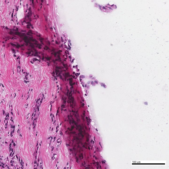
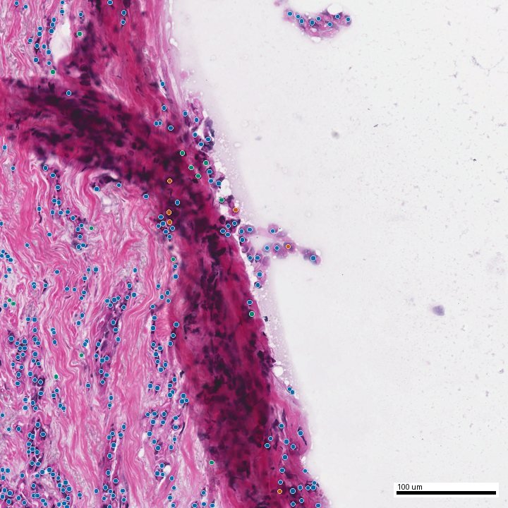
Path23-assisted reportDe-identified sample
H&E tissue field from a de-identified demonstration report
Model-generated nuclei overlay on the same field
H&ENuclei overlay
Drag to compare the tissue with the model-derived overlay
Before you start

Helpful information to gather.

Original laboratory

Name and location of the facility currently holding the slides or block.

Material available

Whether you have a report, glass slides, paraffin block, or existing digital images.

Care-team contact

The treating clinician who should receive and discuss the consultation report.

Do not send any of this medical information by ordinary email. Initial email should contain only your contact details and a request for secure intake instructions.
Request guidance

Start without sending medical details.

Ask for secure intake instructions and tell us only whether the material is digital, glass slides, a block, or still at the original laboratory.

Contact TrueDx

Compose a general enquiry

info@truedx.health

Do not submit patient information. TrueDx is not an emergency service. Discuss treatment decisions with your treating clinician.